|
1.
SM
Home Page
2.
SM Primer Page
3.
SM MRI Comp Page
4.
CM (Chiari Malformation) Comp Page
1. SM Primer Page
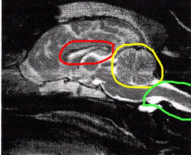
Above is a normal MRI image of a Dalmation
Red circle is normal-sized ventricle in the brain--no
hydrocephalus (fluid in ventricle)
Yellow is normal-sized cerebellum--no squashing or pinching
Green is the beginning of the spinal cord
Extension to the left of the spinal cord is the brain
stem/medulla
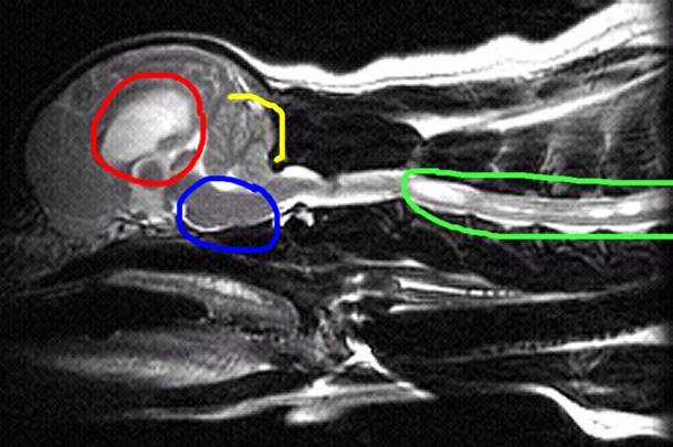
Above is a Cavalier with Syringomyelia (SM)
Red circle is showing enlarged ventricle in brain--hydrocephalus
(fluid inside ventricle)
Yellow is showing a severely squashed/pinched/compressed
cerebellum
Green is showing multiple syrinxes inside the spinal cord, one
quite large (the white is fluid)
Blue is showing a kinked/bent medulla-main part of the brain
stem
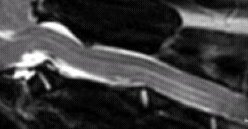
Spinal cord
Faint white line through center of spinal
cord is central spinal canal
Fluid is supposed to flow freely back and forth with
every heart beat inside the spinal cord
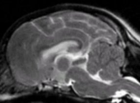
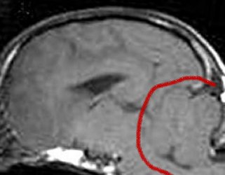
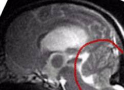
Here is a comparison of squashing/pinching of cerebellum--from
very full to very squashed.
As you look at these you can easily see
the cerebellum is more and more squashed/compressed the further down
you go. There also appears to be a
noticeable shortening of the area behind the main brain in these
particular dogs. If you look at the many MRIs on this
site, you will note that the VAST
majority of Cavaliers are within range of
the 2nd and 3rd ones.
The one at the top is extremely rare in
this breed. It appears almost normal.
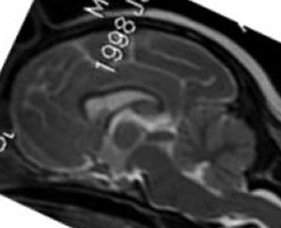
They have done measurements on human chiari
sufferers and a normal control group as well.
The following is a link to a website with this article. It
appears there are significant differences
in the hind skull development between the two groups.
Conquer
Chiari Site
|


