|
1.
SM
Home Page
2.
SM Primer Page
3.
SM MRI Comp Page
4.
CM (Chiari Malformation) Comp Page
3. SM MRI Comp Page
Skull X-Ray to MRI Comps
(including comparison of heads in the 1970s to the late 1990s)
Note: I have left this page mostly as it was when I first did it
in the 1990s. I felt it might still be interesting to some
people. MUCH has been researched since then. There as been a
tremendous influx of new knowledge accumulated over the last 25+
years. We did indeed find out that skull shape has a tremendous
amount to do with whether or not a dog will eventually develop
SM. At the end of this page, I will post links to some of this
newer information and research.
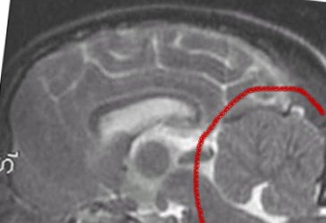
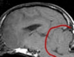
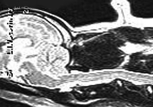
Above is shown a comparison of an MRI showing a full cerebellum
vs. the skull X-RAY of a dog with a full cerebellum and a dog
with an MRI showing a crushed cerebellum vs. the skull X-RAY of
a dog with a crushed cerebellum. The cerebellum is circled in
red. You can easily see there is a huge difference in shape of
cerebellum between these two dogs--the one on the left is quite
full, the one on the right is severely crushed.
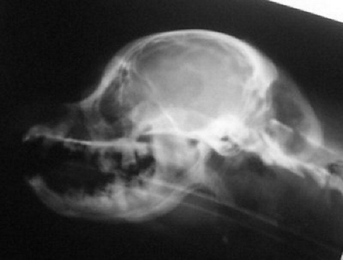
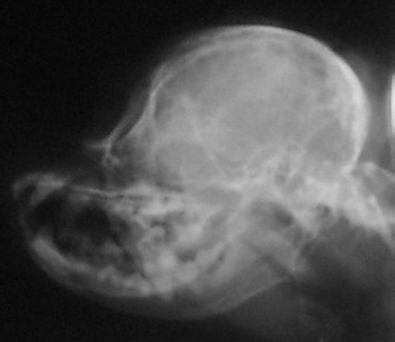
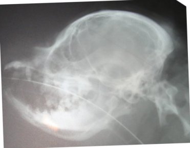
The skull x-ray of a dog with a full cerebellum is below that on
the left and the skull x-ray of a dog with a crushed cerebellum
is shown on the right. You can easily see there is a distinct
difference in the shape of the back of the skull between the two
and why the cerebellum ends up crushed. The most distinct
difference, besides the fact that the skull on the right is
shorter from front to back, is that the occiput sticks out in
the skull x-ray on the left. In the right, that occiput is still
there, but it is more downsloped and blends in with the back of
the skull to the point you can barely see it--almost like
someone took a hammer to the occiput and smashed it in.
Based on my findings, a study was eventually done to determine
if a skull x-ray could be used to determine amount of CM. The
more CM, the more likely there is to be SM--in most cases. This
study did show there was one skull measurement that did indicate
amount of CM. Here is a comparison of two skull x-rays which
show why this particular measurement indicates amount of CM. The
top one is a skull x-ray of a Cavalier with severe CM (and has
SM as well) and the bottom one is a skull x-ray of a Cavalier
with very little to no CM (and no SM).
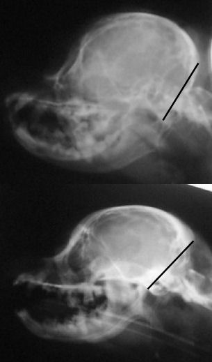
The line was drawn from right in front of the 'hole'
(black spot), center of that curved bone (the 'jaw hinge' area)
to where the most thickened area of the skull ends (and the
occipital area begins). In the dog with almost no CM and no SM
you can CLEARLY see there is a lot more lower backskull left
behind/below that line as compared to the dog above--which
literally has almost none! This also happens to be the area
where the cerebellum sits so it would be common sense that the
dog in the top x-ray would have much less room for the
cerebellum than the dog on the bottom--and therefore more CM (chiari
malformation). There just isn't any skull left for there to be
enough room for the cerebellum. It appears here that a good
skull x-ray may be very useful in determining amount of CM and
therefore breeding towards a goal of less and less CM in future
offspring and thereby lessening the prevalence of SM. A skull
x-ray is much cheaper than an MRI which will enable many more
breeders to x-ray for amount of CM in the future as more
information becomes available.
Below are more side skull x-rays of dogs who have also been MRId.
Their diagnosis will be listed below the comps. The idea is to
see if any differences in skull shape could be ascertained from
a simple skull x-ray. Next to it the MRI was fit into the skull
x-ray, just to see where everything fits in. Below is the
result. So far there appears to be a considerable difference in
the shape of the back of the skull between the dogs with SM and
without!
Below
are Cavaliers WITH SM.

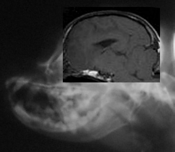
Dog above has moderate to severe malformation and SM with
symptoms
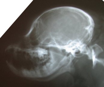
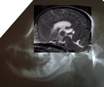
Dog above has moderate malformation and SM with symptoms
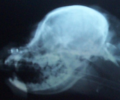
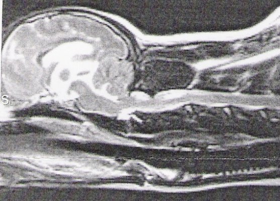
Dog above has moderate malformation, herniation and syrinxes.
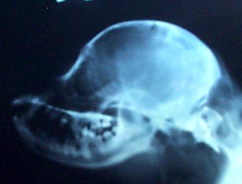
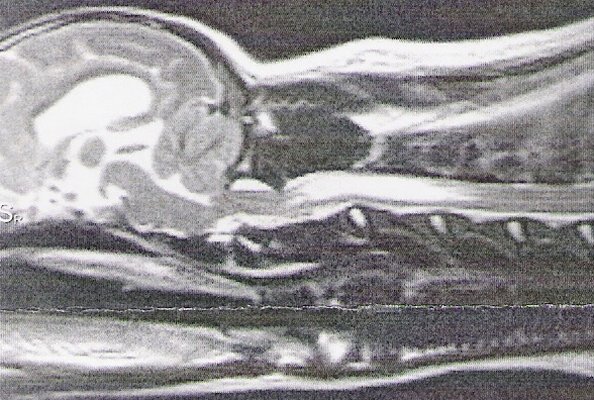
Dog
above has moderate to severe malformation, herniation and
syrinxes.
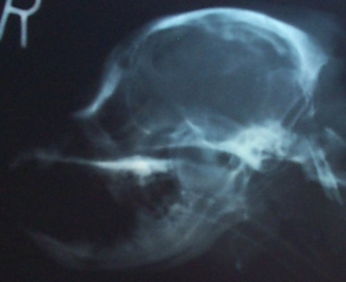
Dog
above has mild malformation, moderate herniation, and syrinxes.
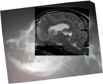
Dog above has moderate malformation and SM with symptoms and has
already has surgery.
You can actually see where the skull was cut away.
Below are Cavaliers without SM.

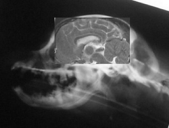
Dog above has extremely mild malformation, no SM, no symptoms
Below are the front and side head pictures of the dog above.

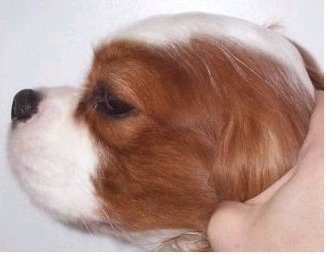
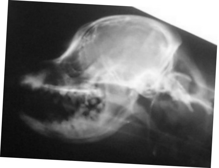
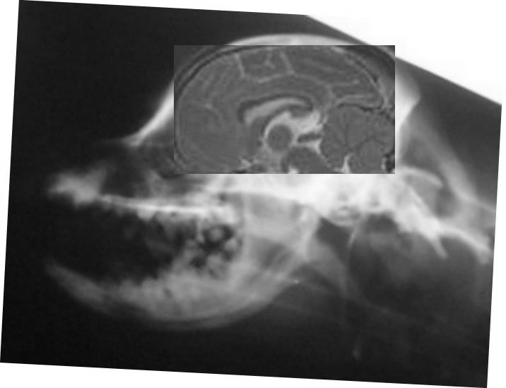
Dog above has extremely mild
malformation, no SM, no symptoms
Below are the front and side head pictures of the dog above.
Sorry his eyes were a bit closed in the front head shot!


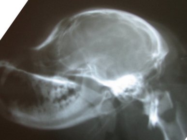
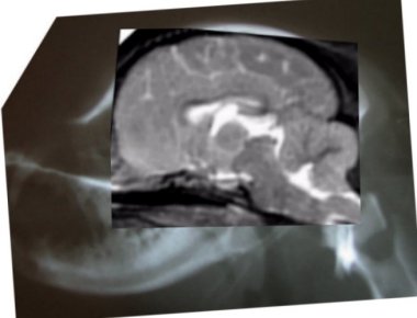
Dog above has mild malformation, no SM, no symptoms
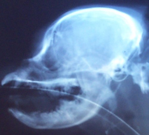
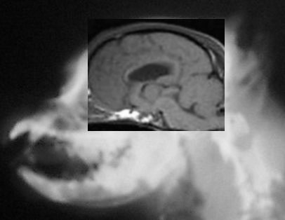
Dog above has mild malformation, no SM, no symptoms
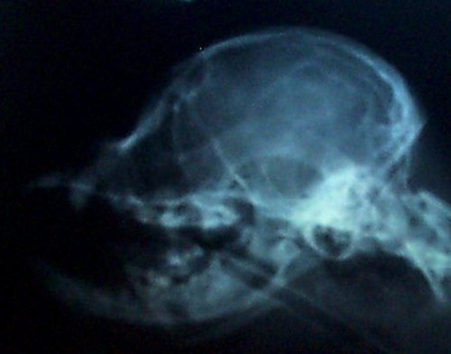

Dog above has mild malformation, no SM, no symptoms
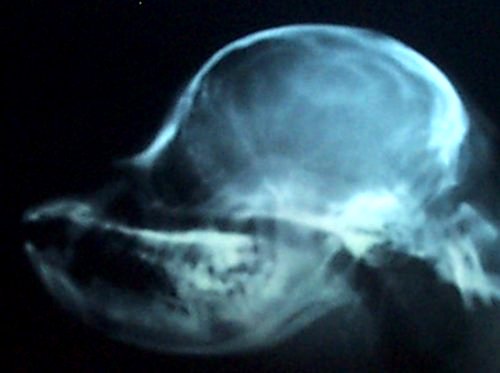
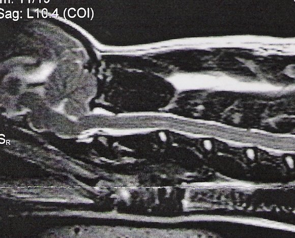
Dog above has mild malformation, no SM, very slight hydromyelia,
no symptoms
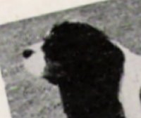
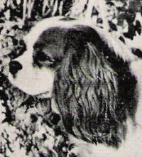
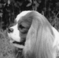
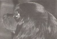
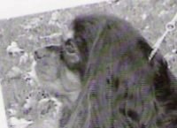
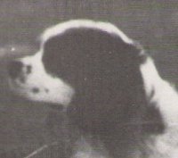
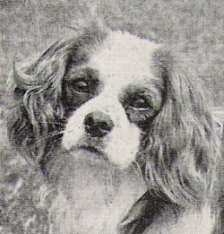
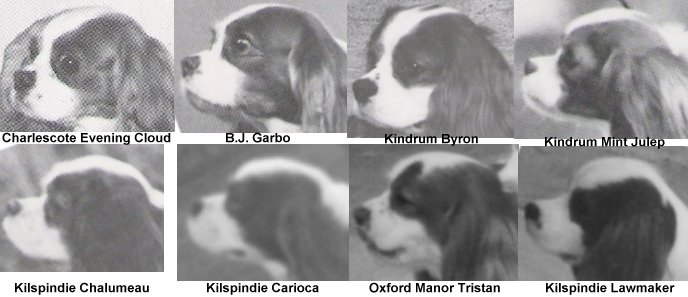
Below are many side head pictures of dogs from the 70s/80s. All
were either imports, had one parent as an import, or all 4
grandparents were imports. You can easily see the longer and
flatter skulls Cavaliers used to have. Most of these also became
CKCSC champions.
Even more head comps--comparing the heads of the 70s to later
1990s. Quite a difference! The skulls in the 70s were more
elongated and flatter whereas today they are much rounder and
more sloped.
Some links to some of the research that has taken place
since the 1990s.
Pilot Study of Head Conformation Changes Over Time in Cavaliers
Video of head changes
|


